How to manage swellings with pus and blood in your livestock

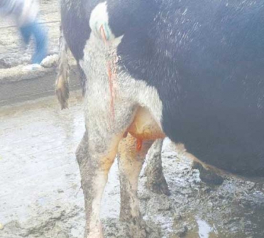
Last week, Vet Perspective carried an article on swellings called hernias. The topic elicited a lot of feedback from readers and that is why we will discuss other conditions that cause swellings in animals.

Abscesses

A collection of pus in a tissue and subsequent swelling in veterinary medical terms is called abscesses, when they occur under the skin they are cutaneous abscess or cellulitis. Abscesses aren’t a reserve for the skin; they can occur in any organ including the brain, liver or lungs and will impair the functioning of these organs. Liver and sole abscesses are common diseases in cattle. Abscesses are also clinical signs for other diseases like lymphadenitis, actinomycosis or lumpy jaw.

These pus containing swellings will present in any shape and anywhere on the animals’ body. The swelling will be painful, soft in consistency and warmer than adjacent body organs.

Abscesses are commonly caused by bacteria. Bacteria will gain access into the tissue through direct entry as occurs during traumatic injuries with contaminated objects. Bacteria can also spread from other infected areas or through blood or lymph from a remote site.

Abscesses may occur at needle injection sites; this occurs when contaminated needles are used to inject drugs or vaccines into the animal’s body. This results in introduction of bacteria at the site and subsequent swelling. In summary, failure to follow strict asepsis and improper administration techniques especially when dealing with drugs that cause tissue irritation can result in abscess.

Management of abscesses

Most superficial abscesses are managed by incisions and draining. All the pus must be drained, and all dead tissues and foreign bodies removed. Now this may not be a simple procedure as many may think and an animal health professional hand is required; in doing all this and sealing of the dead space to prevent reformation of the abscess. When abscesses are poorly managed, they can result in wounds. Always give antibiotics to prevent any secondary infections.

Heamatomas

Hematoma describes blood filled pocket within body tissues. Blood circulates through the body through a series of pipes varying in size and type; if you remember your biology lessons we had veins that carry blood under low pressure back to the heart and arteries that carry blood under high pressure from the heart then there are thin capillaries that perfuse blood through tissues. Now at time these blood vessels rupture and let blood into body tissues and cavities. This leakage results in formation of heamatomas in other words internal heamorrhage or bleeding will cause heamatomas.

They are named according to their location or the affected organs. Common heamatomas in livestock are ear, udder, vaginal and penial heamatomas. They are caused by trauma, bites, rubbing against surfaces and high blood pressure. Excess ear flapping will due to itchiness or head shaking in pigs, dogs and rabbits can cause aural/ear heamatomas. This shaking can be as a result of parasitic infestation.

Udder haematoma is normally caused by calf bites during suckling. Penial haematomas are common in young bulls as a result of the penis bending when erect or in case of repeated mating. Traumatic injuries can rupture blood vessels and cause heamatomas. Signs of heamatomas include, a visible swelling, that appears suddenly, discoloration and itchiness that causes rubbing against objects or head shaking in case of ear heamatomas.

Heamatomas will in most cases resolve by themselves, heparin can be used for the blood dispersion or reabsorption for mild heamatomas. Most heamatomas are treated conservatively if they haven’t resolved themselves after two to three weeks. This is done through aspiration aseptically followed by tight bandaging to prevent fluid from forming. This is followed by an antibiotic cover to prevent any secondary infections.

(Dr. Othieno is a veterinary surgeon working with the Kenya Tsetse and Trypanosomiasis Eradication Council -KENTTEC)