Mastitis detection using California Mastitis Test

Mastitis is a result of the udders' response to microorganisms such as bacteria and fungi invading the milk gland where they rapidly multiply inside it resulting in the changes that are seen.

Types of mastitis

  1. Acute mastitis - it is very severe mastitis. The cow has high fever and stops feeding. Some even die.
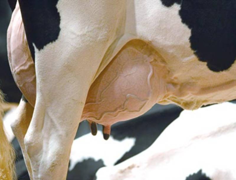
  2. Clinical mastitis - the udder swells up, the skin looks reddish and feels hot. The swollen quarter/teat is painful. The milk looks abnormal with a watery consistency, discoloured (yellow, greenish, cream) and flakes (small aggregates of bacteria/ fungi) may appear in the milk.  The milk flow is considerably reduced or stops completely because teat canals are blocked and secretion of milk by the gland is reduced. Clinical mastitis can be mild, severe or very severe.
  3. Subclinical mastitis: In most cases the beginning of the mastitis after bacteria have entered the milk gland is more gradual. Signs can be so mild as to go almost unnoticed. There may be a very slight udder swelling that disappears quickly. Mild changes in the milk color and consistency that don't last. The flow of milk is not affected and the cow shows no pain and feeds normally. This very common almost invisible form of mastitis is called subclinical mastitis.
  4. Chronic mastitis - subsequent to subclinical mastitis the quarter may return to normal, eliminate all mastitis bacteria from the gland and progress to full recovery. However, a number of cases quarters that look normal remain infected with bacteria for weeks and months. With some mastitis bacteria, the affected quarters can remain infected for the cow’s lifetime. This is called chronic mastitis. Over successive lactations quarters with chronic mastitis change from the normal "spongy" consistency to the hard consistence of dead tissue.

How to detect mastitis using California Mastitis Test?

When the udder is infected by mastitis bacteria, it produces cells of inflammation. A high number of these cells is a clear sign of mastitis. The high number of cells can be detected by examining a milk sample from every quarter.

The most common mastitis detection test, used at milking is the 'California Mastitis Test' (CMT). The CMT fluid is mixed in the ratio 1:1 with the milk (equal amount). The test fluid contains a purple dye and picks up slight invisible changes in the milk that are caused by the mastitis.  If the quarter is affected by mastitis the mix of test fluid and milk becomes thick and may also have a color change. If the quarter is free of mastitis the test fluid-milk-mix does not change color and retains the normal milk fluid consistency.

 How to perform the CMT?

Milk some milk into each cup of the test paddle

The four cups of the test paddle should be maximum half full

Add an equal amount of test fluid to each cup milk: test fluid = 1:1

Mix test fluid and milk by rotating the CMT paddle gently

Where the fluid becomes thick and changes in color the quarter has mastitis

Always remember from which quarter you took the mastitis milk

Interpretation of CMT Results

  • Negative Reaction - the mixture remains watery and does not thicken. It means there is no infection with mastitis bacteria in the quarter.
  • Trace Reaction - there is slight thickening of the mixture, which disappears when paddle is rotated. If all 4 quarters show a trace reaction there is no infection, but if only one quarter shows trace reaction presence of infection in this quarter is possible.
  • Weak Positive Reaction – the mixture clearly thickens but remains liquid (no gel formation). After long rotation of the test paddle (more than 20 seconds) thickening may disappear. This means there is mastitis infection
  • Clear Positive Reaction - the mixture thickens immediately and forms a slight permanent gel, which sits at the center of the test cup when paddle is rotated. There is mastitis infection.
  • Strong Positive – The gel is formed and surface of the mixture remains raised like a fried egg when rotating the paddle. There is severe mastitis infection.

This article has been adapted from Large Animal Medicine textbooks.

Author; Dr. Paul R. N. Kangethe (BVM, UoN)

Email; [email protected]


Want to get latest farming tips and videos?
Join Us